Ines’s Journey: Life after TRAP squence and the loss of her twin
Acardiac twins, otherwise known as twin reversed-arterial perfusion (TRAP) sequence, is a rare and serious complication of monochorionic (one placenta) twins. Although the cause for the syndrome is not completely understood, it has been hypothesized that large vessels on the surface of the common placenta are responsible. Blood is perfused from one twin (“pump” twin) to the other twin (“acardiac” twin) by retrograde (backward) flow. Thus, the acardiac twin receives deoxygenated (oxygen depleted) arterial blood from the pump twin in the wrong direction.
The inadequate perfusion of the acardiac twin is responsible for a spectrum of lethal anomalies, including acardia (absent heart), acephalus (absent skull), severe maldevelopment of the upper body, and a relative excess of edematous (excess fluid) connective tissue. Although the pump twin is structurally normal, there is an increased risk of death (up to 50-75%) for that twin. This is due to two important factors. First, the pump twin’s heart has to work to support the perfusion (pumping of blood)of both the pump twin and the acardiac twin. Eventually, the strain to the pump twin’s heart may be too great, resulting in high-output heart failure. Second, premature delivery or miscarriage may occur due to the polyhydramnios (excess amniotic fluid volume) and/or rapid growth of the acardiac twin.
Risk factors associated with pregnancy loss include polyhydramnios (defined as a maximum vertical pocket of amniotic fluid greater than or equal to 8.0 centimeters), large TRAP twin (estimated fetal weight of the acardiac twin is 50% or greater than that of the pump twin), evidence of heart failure in the pump twin (hydrops), or critically abnormal blood flow patterns identified on Doppler ultrasound. Because of the high risk of pregnancy loss in pregnancies complicated by Acardiac/TRAP sequence in the setting of these risk factors, surgical treatment in the womb to separate the circulatory systems of the twins have been proposed.
Frequency
1 in 35,000 births
Diagnosis
The diagnosis of acardiac twins or TRAP sequence is suggested by the presence of a monochorionic (single placenta) twin pregnancy in which one twin (the pump twin) appears structurally normal (no ultrasound findings consistent with birth defects), while the other twin (the acardiac/TRAP twin) has multiple profound birth defects (as listed in the background section above) which are not compatible with life.
The diagnosis is confirmed with the use of combined pulsed and color Doppler ultrasound studies. This method allows for the documentation of the arterial blood flow perfusing the acardiac/TRAP twin in a retrograde fashion, thus securing the diagnosis.
Once the diagnosis is established, further ultrasound studies must be performed to assess whether that individual pregnancy is in the high-risk category for pregnancy loss. These findings are summarized in the section below titled, “Candidacy for Surgical Treatment”.
Management Options and Outcomes
The following management options and corresponding expected outcomes are listed below for pregnancies complicated by acardiac twins (TRAP sequence) with a high-risk factor, thus meeting criteria for fetal surgery.
1. Expectant management: This means that your pregnancy will be watched closely by frequent ultrasounds and other methods, with the delivery timed to prevent the death of the pump twin in the womb. This is associated with a 50 to 75% risk of pregnancy loss or extreme prematurity.
2. Umbilical cord occlusion: There is approximately a 66% chance that the pump twin will survive, with a 5% risk of neurologic injury.
General Candidacy for Surgical Treatment
The inclusion and exclusion criteria for consideration of surgical intervention to separate the circulatory system of the acardiac twin from the pump twin are listed below. The goal of surgical treatments for TRAP is to stop the flow of blood to the acardiac twin thus relieving the strain on the pump twin.
Inclusion Criteria
All pregnancies must be between 16 and 26 weeks gestation. Once the diagnosis of Acardiac/TRAP sequence has been confirmed, the presence of at least one of the following must be present to be considered a candidate for surgical treatment.
1. Size of acardiac twin exceeds the pump twin (abdominal circumference of acardiac twin larger than that of pump twin)
2. Polyhydramnios (maximum vertical pocket (MVP) > 8cm)
3. Critically abnormal Doppler’s in the pump twin (persistent absent or reversed diastolic flow in the umbilical artery, pulsatile flow in the umbilical vein, and/or reversed flow in the ductus venosus)
4. Fetal hydrops of the pump twin
5. Monochorionic-monoamniotic twins
6. The presence of a short cervix is a relative indication, and will be addressed on an individual basis
Exclusion Criteria
1. Presence of major congenital anomalies of the pump twin
2. Abnormal karyotype (characteristics of cell chromosomes)
3. Ruptured membranes (broken bag of waters)
4. Chorioamnionitis (infection in the womb)
Details of Procedures
Because the peculiarities of each pregnancy complicated by Acardiac/TRAP sequence, it is very important to stress that a single surgical approach is inadequate to provide optimal treatment. Each pregnancy must be individually assessed, and the type of fetal surgery must be tailored to the specifics of each case. Important considerations include surgical access (it is preferable to enter the sac of the acardiac/TRAP twin if possible), the size and position of the acardiac twin, the length of the umbilical cord, and the location and length of the placental vascular connections.
Using the above-mentioned considerations, the following surgical approaches are recommended. Note that most surgeries are performed under local anesthesia with intravenous sedation. About a 2 to 3 millimeter (one tenth of an inch) incision is made on the abdomen to allow the insertion of the microsurgical instruments into the womb. Antibiotics are given to the mother.
Fetal Surgery Techniques
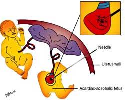
1. Radiofrequency ablation is done with a very thin needle that is inserted where the blood vessels flow into the acardiac twin. Then, guided by ultrasound, a radiofrequency device is used to destroy the blood vessels in order to stop the blood flow. This is all done without any incisions which makes the pain and recovery very similar to an amniocentesis.
2. Umbilical Cord Ligation (UCL): Suture ligature is tied around the umbilical cord of the acardiac/TRAP twin. The procedure could be carried out by ultrasound alone or by combined ultrasound-endoscopy. Some cases do require a second port (incision).
3. Laser Therapy of the Placental Vessels (L-AAVV): Using the techniques originally developed for the treatment of twin-twin transfusion syndrome (TTTS), the communicating vessels on the placental surface are sealed by laser energy.
4. Laser Umbilical Cord Occlusion (L-UCO): The umbilical cord artery then vein is laser occluded using laser energy guided through an operating endoscope.
5. Transection (cutting across) of the umbilical cord of the Acardiac/TRAP twin: This technique is reserved for those cases with monoamniotic twins or where dividing amniorrhexis was performed.
Postoperative Care
Typically, you will remain in the hospital for 1 to 2 days after surgery. You will then be sent home to the care of your primary obstetrician and perinatologist. Weekly ultrasound is recommended for the four weeks after surgery. Then, depending on the clinical circumstances, follow up ultrasounds may be performed every 3 to 4 weeks for the duration of the pregnancy.
Facebook Support Group for Twin Reverses Arterial Perfusion (TRAP) Sequence
Show/Hide Additional Resources
TREATMENT CENTERS
Filter List:
