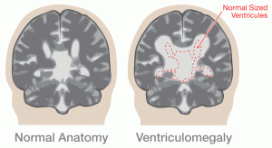
Ventriculomegaly represents enlargement of the fluid collecting system in the brain. It is a pathologic process that has many causes. It may occur due to obstruction of cerebrospinal fluid (CSF) flow, as a consequence of abnormal development of the ventricles or as part of a destructive process as seen in cerebral atrophy. The presence of ventriculomegaly signals an underlying, abnormal central nervous process and is often the first sign of associated abnormalities in organ structures other than the brain. One of the more common causes is due to a narrowing (stenosis) of the tube that connects the 3rd and 4th ventricle (Aqueduct of Sylvius), thereby resulting in the accumulation of CSF. Other common causes involve abnormal brain development such as a Dandy-Walker malformation (abnormal development of the cerebellum) or agenesis of the corpus callosum. Also, chromosomal abnormalities often include ventriculomegaly as one of the physical manifestations.
Frequency
The incidence of isolated fetal ventriculomegaly is 0.5 to 1.5 per 1000 pregnancies.
Diagnosis
Ventriculomegaly is diagnosed prenatally by the presence of enlarged ventricles on ultrasound, though the fetal head measurements may be normal. The lateral ventricles can be visualized as early as 12 weeks of gestation. Several different methods have been proposed to evaluate increased cerebrospinal fluid. The most common method for assessing ventricular size today is to measure the diameter of the lateral ventricle at a location referred to as the atrium. The mean diameter of the normal atrium is 7.6 ± 0.6 mm. Some have suggested that atrial diameters >10 mm indicate the presence of ventriculomegaly. While an atrial diameter of >10 mm is considered abnormal, the term borderline ventriculomegaly is often used to refer to an atrial measurement of between 10 and 12 mm. Borderline ventriculomegaly is associated with an increased risk of central nervous system (CNS) and non-CNS abnormalities, and suggests the need for a more detailed fetal anatomic ultrasound examination searching for other abnormalities. The term hydrocephalus is used when the atrial diameter is > 20 mm.
Management Options and Outcomes

The patient should be referred to center capable of performing a detailed ultrasound of the fetal anatomy due to the high incidence of associated abnormalities in the brain and in other organ structures. Ventriculomegaly will significantly worsen in approximately 2-5% of cases. In many fetuses, particularly those with borderline ventriculomegaly, the condition will resolve spontaneously resulting in a normal outcome. The major factor that influences prognosis is the presence of associated abnormalities. Sometimes a fetal magnetic resonance imaging (MRI) will be performed to further image the brain and other organs. Decisions about management options depend on the gestational age at the time of diagnosis and the presence of associated abnormalities. Studies may be recommended to search for an infectious (cytomegalovirus, toxoplasmosis) or genetic cause (trisomies 9, 13 and 18 as well as triploidy). The decision as to whether to proceed with an attempt at a vaginal delivery versus a cesarean section often depends on whether there is significant head enlargement. If ventriculomegaly is isolated, without significant head enlargement, vaginal delivery is reasonable. If severe abnormalities are present that are incompatible with life, a cephalocentesis (removal of fluid from the ventricular system) may be recommended to reduce the fetal head size, thereby increasing the chances for a successful vaginal delivery.
Candidacy for Fetal Treatment
Historically, in utero, some providers have either performed multiple cephalocenteses (i.e., using an ultrasound-guided needle to remove cerebrospinal fluid [CSF] from the ventricles) or placed a shunt (a tube inserted into the ventricles under ultrasound guidance to drain the CSF from the brain into the amniotic fluid that surrounds the fetus). The goal of these procedures was to prevent progressive damage to the fetal brain caused by the chronically increased CSF pressure. There was a high rate of fetal deaths related to these procedures. Therefore, members of the International Fetal Medicine and Surgery Society (IFMSS) agreed to stop performing these procedures. Currently, there are no in utero procedures being performed on fetuses with ventriculomegaly.
Newborn Care
After stabilizing the newborn, consultation with a medical geneticist and a neurosurgeon is recommended. Postnatal diagnostic imaging of the central nervous system may include computed tomographic (CT) scan and magnetic resonance imaging (MRI). Genetic testing may be performed.
Surgical Management
Once the newborn is stable and an evaluation has occurred, arrangements likely will be made for placement of a ventriculoperitoneal (VP) shunt. Often, if indicated, the goal is to perform this procedure within the first four days of life. In the head, the shunt is usually place in the front portion of the lateral ventricle. The shunt is then tunneled under the skin to the abdominal cavity. The major issues associated with a VP shunt are shunt malfunction and blockage. This complication is quite common resulting in shunt revision in 25% to 50% of cases within the first two years of life. Multiple studies seem to indicate that approximately 40% to 50% of survivors have a normal IQ. The IQs are not proportional to brain expansion after the placement of the shunt. Degree of ventricular dilatation does not appear to be associated with long-term outcome.
Additional Information
Families in whom no genetic cause can be identified should be offered detailed ultrasound evaluation in a subsequent pregnancy due to the 4% recurrence risk.
