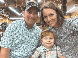
Campbell’s Story: Cincinnati Children’s Offers Expert Treatment for Cloacal Exstrophy
Cloacal exstrophy represents a spectrum of rare anomalies that prevent normal development of a baby’s lower abdominal wall resulting in exposure of the intestines, and bladder. Babies with cloacal exstrophy may also have an imperforate anus, which means the opening to the anus is missing or blocked. There are also a number of other defects associated with this condition, which may require extensive intervention and reconstruction before and after birth.
Because a baby’s organs can be exposed and there is an imperforate anus, this condition can be very life-threatening during and after pregnancy. However, in the past two decades, reconstructive surgery has provided incredible outcomes for babies with cloacal exstrophy.
This is a rare birth defect that affects approximately 1 in every 200,000 to 400,000 births. Babies with the condition are born with a single common channel connecting the rectum, vagina and urinary tract. Part of the large intestine usually develops outside of the abdominal cavity, with the bladder connected to it on either side, in halves. Because the colon is connected to the bladder, urine and stool can mix and there is often no anus. The penis and clitoris are usually split in two and girls may have more than one vaginal opening. Infants born with cloacal exstrophy can also have spinal abnormalities.
What causes it?
Cloacal exstrophy occurs as an isolated event without a recognized cause. There is a higher incidence of cloacal exstrophy in families in which one member is affected as compared with the general population. However, there’s no evidence to suggest that anything done by expectant parents leads to the condition.
Diagnosis
In some cases, cloacal exstrophy is detected from a routine prenatal ultrasound. In other cases, it isn’t diagnosed until birth, when physicians can clearly see the exposed organs. Once diagnosed, additional tests are used to confirm the details of each case and to design a treatment plan. These tests may include:
- Fetal magnetic resonance imaging (MRI)
- Computerized tomography (CT) after the baby is born
- Endoscopy (inserting an instrument to view the inside of an organ)
- Abdominal ultrasound (using sonography to view internal organs and assess blood flow)
Treatment Options
Cloacal exstrophy is treated through an individualized surgical repair after birth, usually in stages to address each defect. This requires an in-depth treatment plan to be created for your child’s specific needs. The extent of cloacal exstrophy surgery required for your baby depends on the type and severity of his or her abnormalities.
In most cases, surgeons perform multiple operations over the course of several years. This approach, referred to as staged reconstruction, usually begins in the first days of life with the highest-priority procedure. Physicians usually repair the bladder, create a colostomy (an opening in the colon with an attached “bag” that allows stool to pass) and repair the abdominal wall defect.
*diagnostic information provided courtesy of our partners at Colorado Fetal Care Center
The Fetal Health Foundation is a parent-founded non profit helping families experiencing a fetal syndrome diagnosis.

