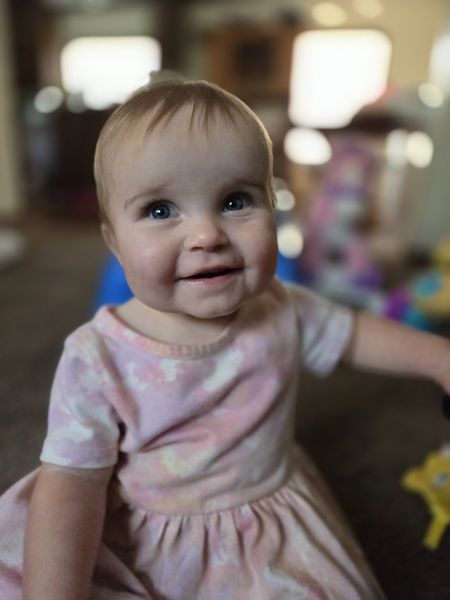
In utero, Lilyana was diagnosed with a tracheoesophageal fistula & esophageal atresia, which can cause breathing, lung, and eating problems. She was born in Cincinnati Children’s Fetal Care Center and had surgery to fix her congenital condition at 2 months old. ❤️ After 96 days in the NICU she is thriving! 👏
Learn about the Aerodigestive & Esophageal Center at Cincinnati Children’s.
What are Esophageal Atresia and Tracheoesophageal Fistula (EA and TEF)?
EA and TEF are two separate and rare conditions that develop in utero but often
occur together.
During the early stages of pregnancy, the trachea (pipe that connects the throat to the
lungs) and the esophagus (pipe that connects the mouth to the stomach) start out as a
single pipe. But as development continues in the womb, a wall typically forms to
separate the two structures: the trachea for air, and the esophagus for food.
Esophageal atresia (EA) occurs when the esophagus forms in two parts (upper and
lower) but doesn’t connect to each other. The result is an inability to properly swallow
food and saliva into the stomach.
Tracheoesophageal fistula (TEF) is marked by abnormal connections in one or more
places between the esophagus and trachea, causing breathing problems.
The causes of TEF and EA are not known, but it is believed there may be a genetic
component.
Some babies with TEF and EA also have other problems with their heart, digestive tract,
kidneys, or bones.
What are symptoms of EA and TEF?
The clinical presentation of TEF depends on the presence or absence of EA. The majority (95%) of TEF cases also have EA.
Symptoms may include:
Difficulty breathing
Coughing or choking when swallowing or trying to eat.
Excessive secretions that cause drooling or frothing from the mouth
Unsuccessful feeding by mouth
Very round belly from trapped gas
Blue skin color
To protect the baby from breathing in (aspirating) their saliva, a small tube may be
placed through the mouth to reach the blind esophageal pouch. This tube is called a
Replogle and helps clear out saliva until surgery can be performed.
How are EA and TEF diagnosed?
Sometimes, using high-resolution ultrasound, babies with EA/TEF can be diagnosed
before birth. Common ultrasound findings include small or absent stomach bubble along
with polyhydramnios (extra fluid in the baby’s sac). Having prior knowledge can help
your care team create a plan to address the conditions once the baby is born.
However, many cases are not diagnosed prenatally and are diagnosed shortly after birth
when symptoms appear. At this point, finding the location of the atresia is important for
avoiding lung damage.
Diagnostic testing includes:
Nasogastric tube and X-ray: a tube is inserted into the baby’s nose or mouth and
down the esophagus. The tube is called a nasogastric tube (NG tube.) When a baby
has EA, the tube hits a blocked end and X-rays can be used to see the location of the
blockage.
The X-rays can also be used to help determine if a TEF is also present. Babies with
only EA do not show gas in the stomach. If the X-ray shows gas in the stomach, this indicates a connection between the trachea and lower end of the esophagus that allows
air to enter the stomach.
Esophagram can be used when a baby may have TEF without EA. In this case, the
baby swallows a contrast fluid (dye) and then X-rays are used to see if the dye enters
the airway. If there is a TEF, the dye is seen passing from the esophagus into the
trachea through a connection.
Rigid bronchoscopy and esophagoscopy uses a telescope and a small camera to
look into the esophagus and trachea to see how far apart the sections of the esophagus
are and if there any connections between the trachea and esophagus.
How are EA and TEF treated?
Surgery soon after birth is required to repair the defects in the esophagus and trachea.
Depending on the location and the baby’s overall health, the type of surgery and
number of surgeries will vary. The goal of the surgery(ies) is to separate the trachea and
esophagus by closing the abnormal channel (fistula) and connecting the two ends of the
esophagus. If the ends of the esophagus are too far apart to connect safely, the
surgeon will delay the repair. In that case, the surgeon will place a feeding tube into the
stomach (gastrostomy tube) and repair the baby’s esophagus in a staged approach
over time.
Additional resources:
Cincinnati Children’s Hospital Medical Center